
Breaking Boundaries Assisting Clinical Diagnosis with Weight-Bearing 3D Imaging

Large FOV
Supports bilateral ankle, knee, and joint
weight-bearing reconstruction imaging
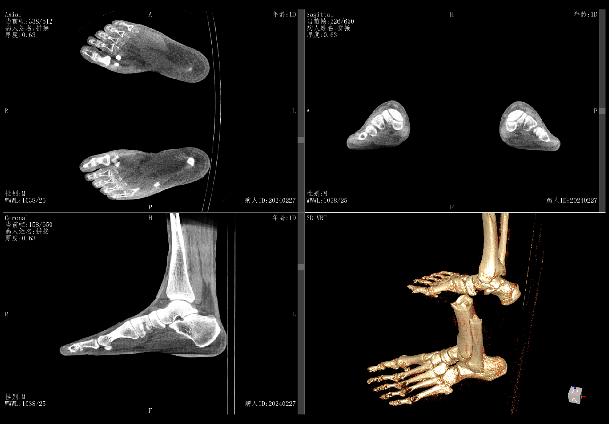

Support MPR and 3D Reconstruction Support post-processing of Radiography, Fluoroscopy and Contrast Imaging
Accurately identifies metal regions and effectively eliminates the impact on image quality
Low-dose acquisition protocols are implemented for different patient sizes, body parts and acquisition modes, effectively lowering radiation dose

Effectively reduces image artifacts
Provides accurate 3D imaging
Meets the examination needs of imaging from skull to ankle
Assists patients with limited mobility in safely and easily getting onto the examination table, enabling all clinical applications of supine X-ray imaging

By utilizing 3D data modeling to identify anatomical features, and through deep learning with big data, precise positioning is achieved, simplifying the workflow of technician.
The LCD touch screen on the system allows for easy access to patient information, positioning guides, and last image reviewing, significantly improving the clinical efficiency.


 Linkedin
Contact Us
010-84575792/3/5/6
international@wandong.com.cn


